How to Understand Anion Gap If Albumin Is Low
What is the anion gap and why does albumin matter?
The anion gap is a derived value used to detect acid-base disorders. It is derived from routine laboratory values in an electrolyte panel, especially serum sodium, serum chloride, and serum bicarbonate. In simple terms, the calculation reflects the gap between measured positive and negative ions in the blood. Because the body maintains electrical neutrality, any gap suggests the presence of unmeasured anions or a shift in the usual balance of charged particles.
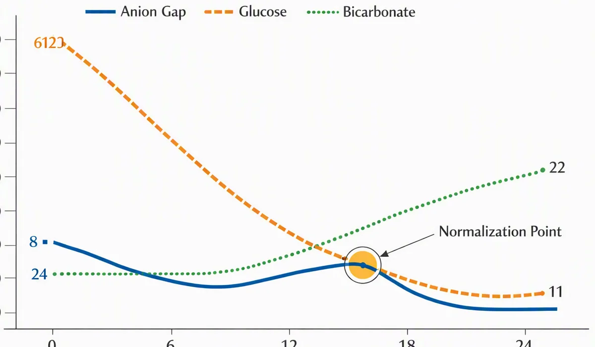
Albumin matters because it is one of the major unmeasured negatively charged serum proteins. When albumin is normal, it contributes significantly to the expected anion gap. When albumin falls, the measured gap can look lower than it truly should, which affects acid-base balance assessment. This is why clinicians pay close attention to albumin when reviewing laboratory values for possible acid-base disorders.
The practical issue is not just the math. A low anion gap can create a false sense of reassurance. If albumin is reduced, a clinically important metabolic problem may be present even when the reported anion gap appears normal. That is where careful interpretation becomes essential for diagnostic accuracy.
How does low albumin change the anion gap result?
Hypoalbuminemia decreases the observed anion gap because albumin is a primary source of unseen negative charge. When albumin declines, the body keeps electrical neutrality by redistributing other ions, often increasing chloride. This means the measured gap can fall even when the patient has a real acid problem. The result is an anion deficit on paper, but not necessarily in physiology.
This matters most when evaluating patients for metabolic acidosis. A patient with low albumin may have a “normal” measured gap while still having high anion gap metabolic acidosis. In that setting, the low albumin has effectively obscured the disturbance, creating what is often called masked acidosis.
The concept of a corrected anion gap helps restore clinical context. It adjusts the observed value upward to account for the loss of albumin-related negative charge. That correction improves clinical significance and helps clinicians avoid missing lactic acidosis, ketoacidosis, or acid retention from renal failure.
In practical terms, a low measured gap does not always mean no problem. If albumin is low, the measured result may underestimate the true burden of unmeasured anions. This is why the corrected value often alters the direction of the workup and the urgency of the assessment.
What’s the albumin correction formula for anion gap?
The most common correction equation adjusts the anion gap for the patient’s blood albumin level. One common method is to add about 2.5 mEq/L to the observed anion gap for every 1 g/dL decrease in albumin below the reference range of about 4.0 g/dL. Correction factors may vary a bit by institution, but the goal is the same: account for the contribution of albumin to the gap.
Standard correction: Corrected anion gap = measured anion gap + 2.5 × (4.0 − serum albumin in g/dL)
As an example, if the measured anion gap is 10 and the albumin is 2.0 g/dL, the correction adds 5.0, giving a corrected anion gap of 15. That change can shift the assessment from apparently normal to clearly abnormal.
The key point is not memorizing one exact equation, but understanding the adjustment factor as a tool for better assessment. The formula estimates how much of the gap is being “lost” because albumin is low. It is an aid to interpretation, not a substitute for clinical judgment.
Keep in mind that the expected normal range for the anion gap itself varies by lab method and whether potassium is included in the calculation. Always interpret the corrected result in the context of the local laboratory’s measurement system and the patient’s full presentation.
When do you use an anion gap calculator?
An Anion Gap Calculator is most useful whenever albumin is abnormal or when the clinical picture suggests an acid-base issue that does not match the measured gap. It can quickly integrate electrolytes and albumin so the clinician can make a more reliable lab interpretation.
You should consider using one when:
- Albumin is decreased and the measured gap appears normal.
- The patient has otherwise unexplained metabolic acidosis.
- The electrolyte panel shows a mismatch between bicarbonate and the anion gap.
- There is suspicion of lactic acidosis, ketoacidosis, or toxin-related illness.
- The patient has chronic illness, inflammation, malnutrition, or renal failure that may affect albumin.
A calculator improves speed and consistency during bedside evaluation. It can also minimize errors when multiple measurements must be reviewed quickly. For clinicians, the main benefit is not convenience alone; it is better diagnostic accuracy. A calculator helps convert the raw numbers into a more reliable clinical picture.
Even so, the calculator should be used as part of a broader process. It does not replace history, physical exam, or blood gas analysis. It is a support tool for careful interpretation, especially when low albumin could otherwise obscure a dangerous metabolic derangement.
How should you read a within-range or high corrected anion gap?
If the corrected anion gap is normal, that points to a major elevated-gap process unlikely, though it still does not completely exclude acid-base disturbance. You still need to look at serum bicarbonate, the patient’s clinical symptoms, and any other signs of compensation. A normal corrected value may align with normal anion gap metabolic acidosis, especially if bicarbonate is anion gap clinical significance low and chloride is elevated.
If the corrected anion gap is raised, that suggests the presence of extra unmeasured anions and raises concern for high anion gap metabolic acidosis. Common causes include lactic acidosis, ketoacidosis, and acid accumulation from renal failure. The correction can reveal a problem that was hidden by hypoalbuminemia.
For example, a patient with low albumin and a measured anion gap of 11 might look reassuring. But after correction, the gap may increase into an abnormal range. That finding changes the clinical significance of the result and should prompt a search for the cause of the acid load. This is exactly where the corrected value prevents masked acidosis from being overlooked.
When the corrected gap is elevated, think in terms of continued acid formation, acid retention, or accumulation of anions that are not directly measured on the standard https://anion-gap-values292.talesignal.com/posts/anion-gap-calculator-for-electrolyte-panel-analysis panel. The decision-making process should then focus on the most likely cause and the urgency of treatment.
What situations can result in a artificially low or within-range anion gap?
A falsely low or unchanged anion gap can arise for multiple causes, and not all of them are due to albumin. The first concern is lab error. Even slight issues in specimen handling, instrument calibration, or reporting can alter the calculation. Because the anion gap is derived from measured values, any error in sodium, chloride, or bicarbonate can affect the result.
Paraproteinemia is another key cause. Abnormal positively charged proteins can lower the apparent gap and cloud the picture. This is one reason a low or surprisingly normal value should not be overlooked without considering the broader clinical context.
Hyperchloremia can also reduce the gap by increasing measured chloride relative to sodium. This may occur in certain fluid shifts, kidney problems, or compensatory states. The result can seem deceptively balanced even when an acid-base disturbance is present.
Other contributors include severe measurement issues, unusual electrolyte patterns, and changes in serum proteins. The takeaway is that a low anion gap is not always benign. It may reflect technical error, altered protein charge, or a real shift in acid-base physiology that calls for further workup.
How is anion gap best interpreted with other acid-base results?
The anion gap should not be interpreted in isolation. It becomes much far meaningful when paired with an arterial blood gas, serum bicarbonate, and the rest of the chemistry panel. In combination, these data help determine whether the patient has acidosis, alkalemia, a compensatory response, or a mixed disorder.
One of the most helpful tools is the delta gap. This calculation compares the change in anion gap with the change in bicarbonate to look for mixed acid-base problems. A mismatch may suggest that more than one disturbance is present, such as high-gap acidosis plus a concurrent metabolic alkalosis or normal-gap acidosis.
The arterial blood gas provides context by showing pH, carbon dioxide, and bicarbonate behavior. If the corrected anion gap is elevated and the blood gas confirms acidemia, the case for metabolic acidosis is more convincing. If the blood gas shows compensation, the clinician can more accurately judge whether the response is appropriate or whether another disturbance is present.
This integrated approach improves diagnostic accuracy. It turns the anion gap from a single number into part of a broader assessment of acid-base status. That matters because low albumin can distort the interpretation unless all the pieces are reviewed together.
Typical questions about anion gap with decreased albumin
The most common clinical question is whether the measured value can be trusted when albumin is low. The response is that the measured value is still real, but its meaning changes. With hypoalbuminemia, the result may underestimate the degree of acid accumulation, so the clinical interpretation should favor correction when acid-base disease is suspected.
Another common issue is whether a patient with low albumin and normal bicarbonate can still have an acid problem. Absolutely, especially if the corrected anion gap is elevated or if other markers suggest a mixed disorder. The diagnostic approach should consider symptoms, trends, and the full electrolyte pattern rather than relying on a single number.
In practice, the best strategy is to combine the corrected gap with the rest of the evaluation. That means reviewing the electrolyte panel, blood gas, and likely causes of acidosis. It additionally means recognizing that the absence of an obvious abnormality on the measured gap does not rule out clinically important disease.
Here are the most common questions and concise answers for quick reference.
How can you correct the anion gap for low albumin?
Use a correction formula that adds about 2.5 mEq/L to the measured anion gap for every 1 g/dL albumin below 4.0 g/dL. This estimates the corrected anion gap and helps account for the effect of low serum albumin on the result.
How does hypoalbuminemia lower the anion gap?
Because albumin is a major negatively charged serum proteins component. When albumin drops, there are fewer unmeasured anions contributing to the gap, so the measured anion gap falls. This is a problem of protein binding and electrical neutrality rather than a true absence of acid-base disturbance.
Can a normal anion gap still hide metabolic acidosis if albumin is low?
Absolutely. Low albumin can create masked acidosis by lowering the measured gap. A patient may still have metabolic acidosis, including high anion gap metabolic acidosis, even if the uncorrected number appears normal.
What is considered a high corrected anion gap?
A raised corrected value is one that exceeds the local lab’s reference range after albumin correction. The exact cutoff depends on the facility and the calculation method, but any rise after correction should be concerning for unmeasured acid load and warrant further assessment.
Should you always adjust anion gap for albumin?
It is not required in every case, but it is especially recommended when albumin is low or when the clinical picture suggests an acid-base imbalance that does not match the measured gap. Using the correction improves assessment, improves better interpretation, and can prevent missed metabolic acidosis from lactic acidosis, ketoacidosis, or renal failure.